“A Field-Cycling Imaging first look at cerebral small vessel disease” was submitted by Dr Nicholas Senn, a Research Fellow at the University of Aberdeen Biomedical Imaging Centre.
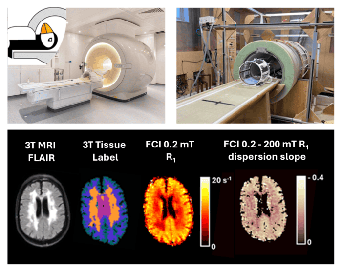
Quantitative maps of relaxation rate (R1 = 1 /T1) at 0.2 mT and rate of dispersion of R1 between 0.2 and 200 mT were generated using data obtained from the mark II Field-Cycling Imaging (FCI) scanner. Depicted alongside these maps are the tissue label map generated from 3T MRI data and the 3T MRI FLAIR image, co-registered to the FCI images. The images shown were obtained from a participant with white matter hyperintensities arising from cerebral small vessel disease.
The PUFFINS 2 study, now nearing the end of participant recruitment, aims to investigate the potential utility of Field-Cycling Imaging for stroke and cerebral small vessel disease. Dr Nicholas Senn said: “From our latest results we observed that measurements of multi-field R1 dispersion obtained from brain and white matter changes associated with cerebral small vessel disease were repeatable between two study visits. The optimal fitting routine, combined with denoising and motion correction approaches, improved the efficacy of the multi-field R1 mapping approach.”
The full paper is now published in Magnetic Resonance Materials in Physics, Biology and Medicine:
Senn, N., Ross, P., Ayde, R., Mallikourti, V., Krishna, A., James, C., de Vries, C.F., Broche L.M., Waiter, G.D., MacLeod, MJ. Field-cycling imaging yields repeatable brain R1 dispersion measurement at fields strengths below 0.2 Tesla with optimal fitting routine. Magn Reson Mater Phy (2025). https://doi.org/10.1007/s10334-025-01230-w